 Before
After
Before
After
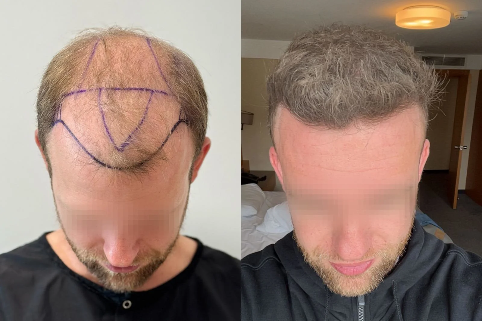
Before surgery, thinning extended across the frontal region and mid-scalp, while a large number of fine, miniaturized native hairs were still present throughout the broader area.
Because the thinning covered a relatively large surface, the possibility of a second session was discussed with the patient before the first procedure.
He had not previously used finasteride or minoxidil. Both were started after surgery.
Case summary
| Clinical detail | This case |
|---|---|
| Norwood stage before surgery | Norwood V |
| Technique | FUE |
| Total grafts | 3,612 |
| Transplanted hair count | At least 7,653 |
| Single-hair grafts | 619 |
| Double-hair grafts | 1,945 |
| Multi-hair grafts | 1,048 |
| Treatment areas | Frontal region and mid-scalp |
| Crown transplanted | No |
| Follow-up shown | 10 months |
| Previous finasteride/minoxidil use | No |
| Finasteride after surgery | Yes |
| Minoxidil after surgery | Yes |
Across our case library, follicular units containing three or more hairs are described as multi-hair grafts.
Treatment planning
Before surgery, the hair loss was classified as Norwood V.
Why was a second session discussed before surgery?
The pre-operative photograph shows thinning across a broad area rather than an isolated frontal recession.
Many native hairs were still present, although a large proportion appeared miniaturized. It was therefore difficult to know before surgery how much additional density the patient might eventually want once the first transplant had matured.
The first procedure focused on the frontal region and mid-scalp, while the possibility of further surgery remained open for later reassessment.
The purple markings visible in the pre-operative photograph show the planned recipient areas before surgery.
Graft composition
The 3,612 transplanted grafts consisted of 619 single-hair grafts, 1,945 double-hair grafts and 1,048 multi-hair grafts.
Based on the recorded composition, the procedure involved at least 7,653 individual hairs:
- 619 single-hair grafts contributed 619 hairs
- 1,945 double-hair grafts contributed 3,890 hairs
- 1,048 multi-hair grafts contributed at least 3,144 hairs
The true number may have been higher because our multi-hair category includes follicular units containing three or more hairs.
Across our case library, we do not assign exact retrospective graft counts to the frontal region, mid-scalp or crown when treatment-zone boundaries overlap. Doing so would imply a level of precision that the surgical distribution does not support.
Was the crown transplanted?
No. The crown also showed thinning but was not grafted in this session; all 3,612 grafts went to the frontal region and mid-scalp, where the first session was concentrated.
The 10-month result
At 10 months, the follow-up photograph shows greater visible coverage across the frontal region and the visible part of the mid-scalp.
Both photographs show the head tilted forward and expose a meaningful portion of the top of the scalp, making the broader pattern reasonably useful for comparison.
There are still differences between the images. The follow-up hair is longer and styled differently, while the exact head position is not identical.
The hair also appears lighter and more ash-toned in the follow-up photograph. From the images alone, we cannot determine whether this comes from lighting, hair color or a combination of both. Lower contrast between the hair and scalp can make scalp show-through less noticeable, so the color difference should be considered when comparing coverage.
The photographs document the cosmetic change but cannot provide an exact percentage increase in density or graft-survival rate.
The image records the result at 10 months, with further maturation still possible.
Follow-up of the miniaturized areas
Before surgery, numerous fine, miniaturized hairs were visible within the broader thinning area.
The patient reported using finasteride and minoxidil consistently after transplantation.
At the 10-month clinical follow-up, coverage in areas that had contained many miniaturized hairs appeared improved. Transplanted grafts had also been placed within the broader treatment area, so the available photographs and routine clinical follow-up cannot determine exactly how much of that change came from transplanted growth and how much came from pre-existing native hair.
Why was the second session deferred?
At the 10-month follow-up, the patient’s overall coverage had improved enough that he did not feel an immediate need for additional density.
The option of a second session was therefore left open for the future instead of being scheduled at that stage.
Further surgery can be reconsidered if his native hair changes over time or if he later wants additional coverage.
Finasteride and minoxidil after surgery
The patient had not used either treatment before transplantation and started both afterward.
Their use is relevant clinical context because native hair remained throughout the treated areas.
For a more detailed discussion of post-operative medical treatment and native-hair changes, see our 3,853-graft FUE case with post-operative finasteride and minoxidil.
What this case shows
A second session was on the table before the first one began. The patient had thinning over a broad area and a great deal of fine, miniaturized hair, and the first procedure used 3,612 grafts, carrying at least 7,653 hairs (619 single, 1,945 double and 1,048 multi-hair units), across the frontal region and mid-scalp.
At 10 months there was no reason to schedule it. The patient had used finasteride and minoxidil consistently, and the areas that had been full of miniaturized hair looked stronger, although the photographs cannot show how much of that came from the grafts and how much from his own hair. The second session remains an option rather than a plan.
Dr. Mesut Demir’s note
“He had a large number of miniaturized hairs before surgery, so we explained from the beginning that a second session might eventually be useful.
He told us he had been consistent with finasteride and minoxidil after the transplant. At follow-up, the overall response was better than I would usually expect in a case that looked like this before surgery, so there was no need to decide on a second operation at that point.
We can reassess the area later if his hair changes or if he wants additional density.”
Dr. Mesut Demir · Pure Line, Istanbul
About this result
This page documents an individual patient’s result at 10 months.
Individual results vary with donor capacity, hair characteristics, hair loss progression, healing and treatment.
The photographs document this patient’s outcome and should not be interpreted as a guarantee that another patient will achieve the same result.
The patient gave written consent for these photographs to be published.
Information about medication on this page describes this individual’s treatment history and is not a recommendation or prescription for another patient. Medication decisions should be made individually with an appropriate clinician.
Medically reviewed by Dr. Mesut Demir, M.D. ·
What is realistic for your case?
Send clear photos of your hairline, crown, and donor area. We will review what can be planned safely and whether surgery makes sense at this stage.
Review My Case