
Quick answer
Crown results often look slower than frontal results, but the evidence does not show that vertex grafts follow a different biological growth cycle. Transplanted hairs commonly shed in the first weeks, new growth becomes visible after a lag of roughly three months, and appreciable growth is often reported around 6–9 months, with caliber and cosmetic overlap continuing to improve afterward. The crown is simply harder to judge: hair radiates through a whorl across a curved, overhead-lit surface, so growth and visual coverage are not the same endpoint. It is also expensive in donor terms — at the 30–40 follicular units per cm² described in one FUE series, an 8 cm circular area of roughly 50 cm² would represent about 1,500–2,000 grafts before accounting for existing hair. Because donor supply is finite and androgenetic alopecia is progressive, Dr. Mesut Demir decides the vertex from donor reserve, age and likely future pattern rather than from the size of the bald spot alone.
The crown is one of the most misunderstood areas in hair transplantation.
Part of the problem is simple: you can't properly see your own crown.
A patient can inspect a frontal hairline every morning from the same distance in the same mirror. The vertex is different. Most people judge it from a phone held above the head, a second mirror, a barber's comment, or somebody else's photograph. Change the ceiling light, camera height, flash or hair length and the same crown can look like four different crowns.
I see this in follow-up all the time. A patient who feels reassured by his frontal result sends an anxious crown photo and says that it looks much thinner in the office than it does at home. Often, both photographs are telling the truth. The lighting has changed what the scalp reveals.
That psychological detail matters because the crown is the one major transplant area the patient usually evaluates indirectly. It helps explain why patience is harder there.
- Why does the crown look different from the hairline?
- Does crown hair actually grow more slowly?
- How many grafts can a crown hair transplant need?
- Why can the crown still look thin after growth starts?
- Should the hairline or crown be treated first?
- Why does age matter when planning a crown transplant?
- Can medication protect the crown?
- Do PRP or LLLT make crown growth faster?
- When should crown results be judged?
- Can shock loss happen after a crown transplant?
- How I plan the crown
- Frequently asked questions
Why does the crown look different from the hairline?
The frontal hairline frames the face. A relatively modest change in the front can alter how the entire face is perceived.
The crown doesn't have that advantage. It sits on a curved surface and its hairs rotate around a whorl. Overhead light can pass between hairs and reflect directly from the scalp. Wet hair, short hair and high hair-to-skin color contrast make that effect stronger.
The result is a strange but important distinction: hair growth and visual coverage aren't the same endpoint.
A crown may contain growing transplanted hairs and still look disappointing in a harsh photograph. This doesn't prove that graft survival is poor. It may simply mean that the hairs aren't yet long or thick enough to overlap effectively around the whorl.
| Planning factor | Frontal hairline | Crown / vertex |
|---|---|---|
| Main visual role | Frames the face | Covers a top-facing curved surface |
| Hair direction | Mostly directional with controlled transitions | Rotates around one or more whorl centers |
| Visual return per graft | Often high | Often lower over a large area |
| Light exposure | Less direct from above | Frequently direct overhead light |
| Patient can inspect it easily | Yes | Usually no |
| Risk if future loss progresses | Hairline can become isolated | Transplanted island can become exposed as surrounding native hair miniaturizes |
| Donor strategy | Usually high priority | Often secondary to frontal framing in limited donors |
| Second session | Sometimes | More commonly worth discussing when the area is large or future loss is uncertain |
Does crown hair actually grow more slowly?
This deserves a more careful answer than the usual clinic slogan.
After transplantation, many grafted hairs shed during the first few weeks. Published surgical reviews describe a lag of roughly three months before new growth becomes visible, and an observational FUE study reported appreciable growth at around 6–9 months. [9,10] A small follicular-unit survival study that counted transplanted hairs at 6 and 12 months found similar survival at those two later time points, which is another reminder that early appearance and eventual survival aren't the same thing. [11]
What the literature doesn't establish very well is that a vertex follicle has a fundamentally slower biological clock simply because it was implanted into the crown.
So I explain it differently to patients: the crown often looks slower.
The whorl spreads hairs in multiple directions. The recipient surface can be large. Light comes from above. Existing native hairs may still be miniaturizing. Those factors delay the moment when the area looks cosmetically convincing, even if growth is progressing normally.
That is a more defensible explanation than pretending every crown has its own special 18-month biology.
How many grafts can a crown hair transplant need?
There isn't one honest number.
A graft estimate depends on crown surface area, existing native hair, hair caliber, hair-to-skin contrast, curl, whorl geometry, the safe donor supply and how much density we are trying to create.
But some arithmetic helps show why a crown can become expensive in donor terms.
A retrospective FUE study described roughly 30–40 follicular units per cm² as a density associated with a good cosmetic result in its series. [9] That isn't a universal prescription and I don't simply multiply every bald area by 40. Still, it is useful for scale.
If we pretend for a moment that a crown is a perfect circle:
| Approximate crown diameter | Surface area | At 30 FU/cm² | At 40 FU/cm² |
|---|---|---|---|
| 6 cm | 28.3 cm² | ~850 grafts | ~1,130 grafts |
| 8 cm | 50.3 cm² | ~1,510 grafts | ~2,010 grafts |
| 10 cm | 78.5 cm² | ~2,355 grafts | ~3,140 grafts |
Real crowns aren't perfect circles, they contain variable amounts of native hair, and I don't implant every square centimeter at a uniform density. The table is an illustration, not a quotation for surgery.
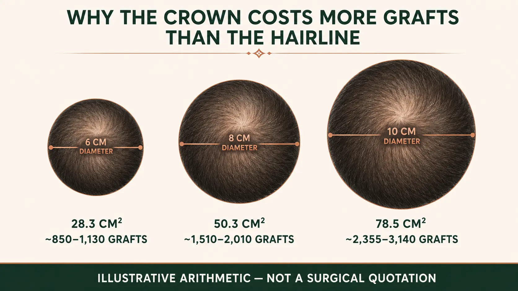
It does explain something patients often underestimate: increasing the diameter of a circular thinning area increases its surface area, not just its width. A crown that looks only a little wider can require many more grafts.
The donor side of the equation
Hair transplantation redistributes a limited resource.
Practice guidelines recommend harvesting only within a safe donor area and describe that area as roughly 189–203 cm², while emphasizing that no single boundary is safe for every patient. [1] The same guideline warns against overharvesting and gives a conservative single-pass example of about 10–15 excisions per cm² in an average-density donor. [1]
A study of 580 men found a mean scalp donor density of 78.2 follicular units/cm² in that population. Using its own 25% extraction model, the estimated first-pass availability varied from about 2,064 FUs in a limited donor zone to 3,612 in an extended zone. [8] Those numbers shouldn't be transplanted from a study table into an individual patient's plan. Ethnicity, donor dimensions, miniaturization and future loss change the answer.
But they make the principle concrete: a crown can consume a meaningful fraction of what a patient has available for life.
I don't start a consultation by asking how many grafts I can put into the vertex. I start by asking how many grafts I can spend there without regretting the decision later.
For the broader logic behind that decision, I have written separately about long-term hair loss planning.
Why can the crown still look thin after growth starts?
Graft count is only one part of density.
Hair caliber can change the visual result dramatically. A 2022 measurement study sampled 270 donor hairs from three men and found an average manual-caliper diameter of 53.3 µm. Scanning electron microscopy showed the hair shaft was elliptical, with mean long and short axes of 83.0 µm and 51.5 µm. [7] It was a tiny sample, so I wouldn't call those universal human averages. What it demonstrates well is that “thick hair” isn't just a casual description. Caliber is measurable, and it matters in surgical planning.
Coverage is also affected by:
- hair-to-skin color contrast
- curl or wave
- the number of hairs within each follicular unit
- crown diameter
- existing miniaturized native hair
- the direction and number of whorls
- hair length
- lighting
- future progression of androgenetic alopecia
A patient with coarse, wavy hair and low color contrast may create much stronger visual coverage per graft than a patient with fine, straight, dark hair over pale skin.
Biology refuses to use the same spreadsheet for everybody.
Should the hairline or crown be treated first?
When donor supply is limited, I usually give the frontal frame more weight.
That doesn't mean the crown is unimportant. It means the front often produces more visual change per graft and is harder to ignore if future loss progresses. In an advanced pattern, using thousands of grafts to make the crown look impressive today can leave too little reserve for the midscalp or frontal zone later.
Hair-transplant guidelines make the same broader point from the donor side: safe-donor boundaries, current miniaturization, family history and likely progression should be assessed before harvesting. [1]
I therefore think about three questions together:
- What bothers the patient most now?
- What pattern is likely to exist years from now?
- What donor reserve will remain after today's operation?
Depending on the case, I may balance the front and crown in one session, stage the crown for later, use a lighter central-whorl strategy, or leave it alone while we try to stabilize native hair medically.
Leaving the crown alone is not very exciting to sell. It can still be the better operation plan.
Why does age matter when planning a crown transplant?
Age matters because the crown I see today may not be the crown the patient has ten or twenty years from now.
Androgenetic alopecia is progressive. Hair-transplant practice guidelines therefore treat age as an important part of case selection and recommend particular caution in younger patients because future loss can undermine an operation that looked reasonable at the time. [1] The same guidelines include the patient's age and the possibility of future sessions among the factors that determine how many grafts can safely be harvested over time. [1]
A 25-year-old with early vertex thinning and a 45-year-old with a similar-looking crown are not automatically the same surgical problem. The difference isn't the birthday itself. It is how much of the future pattern is visible.
At 25, the crown may still be expanding while the frontal and midscalp pattern is also evolving. If I spend heavily on the vertex before that pattern declares itself, I can create a dense transplanted center surrounded years later by thinning native hair, while having fewer donor grafts left to repair the areas that matter more visually. In a younger patient, stabilization with medical treatment and serial photographs may therefore be more valuable than rushing to fill the crown.
At 45, I often have a longer natural history to examine. If the pattern has been relatively stable, the donor remains strong and the frontal priorities are already addressed, I can make a more informed decision about how much donor the crown deserves. That still doesn't make every older patient a good candidate; a rapidly progressing 45-year-old with weak donor hair can be a worse candidate than a well-stabilized younger patient.
So I don't use an age cutoff as an automatic yes-or-no rule. I use age as one part of a larger question: how predictable is this patient's future hair-loss map?
That is especially important in the crown because a vertex can absorb a large graft budget before the final extent of hair loss is clear.
Can medication protect the crown?
Yes, in suitable patients, especially when the vertex still contains miniaturized native hairs.
This is important because a transplant replaces coverage in selected areas; it doesn't make the surrounding native follicles resistant to androgenetic alopecia.
Finasteride
In two pivotal trials, 1,553 men aged 18–41 received finasteride 1 mg daily or placebo. Hair counts were measured in a 1-inch-diameter vertex area, 5.1 cm². Compared with placebo, the finasteride group showed a net advantage of 107 hairs at one year and 138 hairs at two years in that target area. [2]
That doesn't mean every crown gains 107 hairs. It shows that the vertex was directly studied and that finasteride can materially affect the native-hair side of the equation.
I discuss the mechanism, dosing evidence and realistic expectations in more detail in my finasteride guide.
Topical minoxidil
In a 48-week randomized trial of 393 men, 5% topical minoxidil produced 45% more hair regrowth than 2% minoxidil by target-area hair counts and performed better than placebo. [3] The study enrolled men with vertex-pattern androgenetic alopecia, so the evidence is directly relevant to crown thinning.
Oral minoxidil
Oral minoxidil shouldn't be treated as an automatically stronger version of topical minoxidil.
A 2024 double-blind randomized trial enrolled 90 men with Norwood 3V–5V loss and compared oral minoxidil 5 mg once daily with topical minoxidil 5% twice daily for 24 weeks. Oral treatment didn't demonstrate overall superiority for the primary density outcomes, although photographic assessment of the vertex favored the oral group. [4]
Oral minoxidil can be useful in selected patients, but it is a medical prescription with systemic adverse effects to consider. Treatment should be individualized rather than copied from somebody else's protocol online.
Do PRP or LLLT make crown growth faster?
Supportive treatments may improve androgenetic alopecia in some patients, but that is different from proving that transplanted crown grafts will mature faster. Neither PRP nor LLLT can guarantee a shorter post-transplant timeline.
PRP
A meta-analysis of 10 randomized trials, representing 555 treatment units, found an average hair-density advantage of 25.09 hairs/cm² for PRP versus control. It did not find a statistically significant improvement in hair diameter, and smaller studies tended to report larger density effects. [5] A newer systematic review also described substantial heterogeneity and limited certainty across PRP protocols. [6]
That is useful evidence, but it is not permission to promise a “faster crown.” PRP preparation, platelet concentration, injection schedules and study designs vary considerably.
Low-level light / laser therapy
A 2019 meta-analysis included eight studies comprising 11 double-blind randomized trials and found a significant increase in hair density with LLLT versus sham treatment. [12] Again, that concerns androgenetic alopecia treatment. It doesn't prove that a laser cap can turn a normal post-transplant timeline into a three-month final result.
Supportive treatments may have a place. I don't use them as an excuse for poor donor planning or unrealistic timelines.
When should crown results be judged?
I prefer ranges to false precision.
Published hair-transplant literature describes shedding in the first weeks, new growth beginning after a lag of roughly three months, and appreciable growth around 6–9 months. [9,10] Some series assess final or near-final outcomes at 12 months or later. [11]
In practical follow-up, I explain the crown like this:
- Weeks 2–4: transplanted shafts commonly shed
- Months 2–3: the area can look frustratingly similar to baseline
- Months 3–6: new growth begins to become visible, but hairs may be fine and uneven
- Months 6–9: the cosmetic change becomes much easier to judge
- Months 9–12: caliber, length and overlap continue improving
- After 12 months: some patients still notice further cosmetic maturation, particularly when the crown needed a large amount of visual coverage
I don't use “15–18 months” as a magic deadline. Some patients continue to improve late, but a weak result should not be dismissed indefinitely with “the crown is slow.” If the appearance is genuinely concerning, I compare standardized photographs, examine growth, review scalp health and look at the surrounding native hair.
One of the worst habits in this field is using patience as a substitute for assessment.
A crown photograph is only useful if the photograph is comparable
For follow-up, I prefer the same:
- room or light source
- camera angle and distance
- dry or wet hair state
- hair length where possible
- no flash unless flash was used in the baseline image
If the 6-month image is taken under a bathroom spotlight and the 9-month image beside a window, the comparison can tell you more about lighting than hair.
Can shock loss happen after a crown transplant?
Yes.
Recipient-site effluvium, usually called shock loss, can affect native hair within or around a transplanted area. Reviews describe onset commonly within a few weeks after surgery, with advanced miniaturization increasing concern. Most cases are temporary, but very weakened native follicles may not recover fully. [13]
This is particularly relevant in a crown that isn't completely bald. Dense placement among fragile miniaturized hairs isn't the same technical situation as implanting into smooth, empty scalp.
It is one reason I want to know what hair is still alive before deciding how aggressively to operate.
How I plan the crown
I don't start with a graft number.
I look at the donor area, caliber, density, miniaturization, age, current Norwood pattern, family history, medication history, crown diameter, whorl direction, frontal priorities and the amount of future loss the donor may need to cover.
Scalp health also matters. Active inflammation should be understood before an elective transplant; I cover that separately in my guide to seborrheic dermatitis and hair transplantation.
Then I decide whether today's best plan is to:
- transplant the crown fully
- use a lighter central-whorl strategy
- split grafts between front and crown
- prioritize the front and reserve the crown for a second session
- treat medically and reassess
- or protect the donor and leave the crown alone
I have told patients not to spend grafts on the crown. That is occasionally the most useful thing I can do in a crown consultation.
I would rather explain a limitation before surgery than invent an explanation after it.
Is a second crown session sometimes necessary?
Yes, and discussing that possibility before the first operation is healthier than presenting it later as a surprise.
A large vertex may simply have more surface area than can responsibly be covered at the desired density in one session. In other patients, I deliberately preserve donor reserve because the surrounding pattern is still evolving. If the first session matures well and the donor remains suitable, a second procedure may be considered for additional coverage.
That doesn't mean crown transplantation inherently requires two surgeries. It means one-session density should never be planned as if donor hair were unlimited.
Final thought
The crown can improve beautifully after hair transplantation, but it rewards restraint.
It is difficult to photograph consistently, visually expensive in grafts and unusually dependent on caliber, contrast, hair direction and future loss. The patient also has the peculiar disadvantage of not being able to look at it naturally in a mirror.
So I don't treat the crown as an empty circle waiting to be filled.
I treat it as one part of a long-term map.
The operation has to make sense when the grafts grow. More importantly, it should still make sense if the native hair around them keeps changing.
Frequently Asked Questions
Why does the crown look slower after a hair transplant?
Is slow crown growth always a bad sign?
When does crown hair usually start growing?
How many grafts does a crown hair transplant need?
Is a crown hair transplant worth it?
Should I transplant the crown or the front first?
Is a crown transplant a good idea in a young patient?
Can finasteride help crown hair?
Can minoxidil help the crown?
Does PRP speed up crown transplant growth?
Does a crown transplant need a second session?
Can shock loss happen after crown transplantation?
Why does my crown look different in every photo?
Can a crown be made as dense as it was before hair loss?
— Dr. Mesut Demir
Wondering whether your crown is worth the grafts?
Send us clear photographs of the crown, hairline, mid-scalp and donor area along with your age, family history and any treatments you are already using. We will tell you what the pattern suggests, how much donor the vertex would realistically cost, and whether the crown is the right place to spend it now.
Request a Case Review- Mysore V, Kumaresan M, Garg A, et al. Hair transplant practice guidelines. Journal of Cutaneous and Aesthetic Surgery. 2021;14(3):265–284. doi:10.4103/JCAS.JCAS_104_20
- Kaufman KD, Olsen EA, Whiting D, et al. Finasteride in the treatment of men with androgenetic alopecia. Journal of the American Academy of Dermatology. 1998;39(4 Pt 1):578–589. doi:10.1016/s0190-9622(98)70007-6
- Olsen EA, Dunlap FE, Funicella T, et al. A randomized clinical trial of 5% topical minoxidil versus 2% topical minoxidil and placebo in the treatment of androgenetic alopecia in men. Journal of the American Academy of Dermatology. 2002;47(3):377–385. doi:10.1067/mjd.2002.124088
- Penha MA, Miot HA, Kasprzak M, Müller Ramos P. Oral minoxidil vs topical minoxidil for male androgenetic alopecia: a randomized clinical trial. JAMA Dermatology. 2024;160(6):600–605. doi:10.1001/jamadermatol.2024.0284
- Effectiveness of platelet-rich plasma in the treatment of androgenic alopecia: a meta-analysis. Aesthetic Plastic Surgery. 2024;48(5):977–984. doi:10.1007/s00266-023-03603-9
- The role of platelet-rich plasma in androgenetic alopecia: a systematic review. Journal of Cosmetic Dermatology. 2024;23(5):1551–1559. doi:10.1111/jocd.16185
- Bayramoglu A, et al. Hair diameter measurements for planning follicular unit extraction surgery (FUE): is there a correlation between the micrometer caliper and scanning electron microscopy findings? Journal of Cosmetic Dermatology. 2022;21(3):1086–1092. doi:10.1111/jocd.14185
- Chouhan K, Kota RS, Kumar A, Gupta J. Assessment of safe donor zone of scalp and beard for follicular unit extraction in Indian men: a study of 580 cases. Journal of Cutaneous and Aesthetic Surgery. 2019;12(1):31–35. doi:10.4103/JCAS.JCAS_142_18
- Hair transplantation by follicular unit extraction for male androgenetic alopecia: a retrospective observational study from two centers. Medical Journal, Armed Forces India. 2020;76(4):430–437. doi:10.1016/j.mjafi.2019.11.001
- Romera de Blas C, Vega Díez D, Ricart Vayá JM, Gómez Zubiaur A. Complications in follicular unit excision hair transplantation: current evidence and practical approaches. Frontiers in Medicine. 2026;13:1750989. doi:10.3389/fmed.2026.1750989
- Evaluation of survival rate after follicular unit transplantation using the KNU implanter. Dermatologic Surgery. 2001;27(8):716–720. doi:10.1046/j.1524-4725.2001.01029.x
- Liu KH, Liu D, Chen YT, Chin SY. Comparative effectiveness of low-level laser therapy for adult androgenic alopecia: a systematic review and meta-analysis of randomized controlled trials. Lasers in Medical Science. 2019;34(6):1063–1069. doi:10.1007/s10103-019-02723-6
- Garg AK, Garg S. Complications of hair transplant procedures — causes and management. Indian Journal of Plastic Surgery. 2021;54(4):477–482. doi:10.1055/s-0041-1739255