
Quick answer
The Norwood Scale classifies the visible pattern of male androgenetic alopecia from Type I to Type VII, with additional Vertex and Type A variants. Type III is generally the first category regarded as established balding, but a Norwood number is only a description: it does not diagnose the cause, measure donor supply or show how quickly the loss is moving. For hair transplantation the combination matters more than the number — a 24-year-old at Norwood III, a 45-year-old at Norwood III and a 24-year-old at Norwood III Vertex may need three different recommendations. The scale is also an imprecise instrument. Trained observers agreed exactly 85.4% of the time in one study while men grading themselves matched observers only 48.5–55.8% of the time, and a separate study found unsatisfactory inter-rater agreement across the full scale. Dr. Mesut Demir therefore treats the stage as a shared description and plans from age, serial photographs, miniaturization, donor measurements, hair caliber, scalp-to-hair contrast and scalp health.
A common consultation starts with a patient saying, “I think I'm a Norwood IV.”
He has already compared his hair to five diagrams, taken a photograph under a ceiling light and checked his father's and both grandfathers' hairlines.
Then I see the crown view and the photograph from three years earlier. Sometimes the apparent IV is a III Vertex exaggerated by lighting. Sometimes the patient has called himself a II because the front still styles well, while the old photograph shows clear crown and mid-scalp progression.
The label changes because the viewpoint and time frame change. That is worth understanding before the number is used to plan surgery.
- What the Norwood Scale actually measures
- Where the scale came from
- How common is each stage by age?
- Norwood stages I to VII
- Norwood III vs III Vertex vs IIIA
- The age-stage planning matrix
- What the scale leaves out
- Why people often grade themselves incorrectly
- When BASP is more useful
- How I plan surgery beyond the number
- When another condition resembles a Norwood pattern
- Frequently asked questions
What the Norwood Scale actually measures
The scale is a visual classification of patterned loss across three regions: the frontotemporal corners, the frontal and mid-scalp, and the vertex, or crown.
It helps clinicians describe extent and distribution. It doesn't measure miniaturization, establish stability, count the donor area or confirm that androgenetic alopecia is the correct diagnosis.
That distinction matters. A patient can appear to be Norwood II in a photograph while magnified examination shows active miniaturization extending well behind the visible hairline. Another patient can have a naturally high or mature hairline that has barely changed for years. The outline can look similar; the biology isn't necessarily the same.
Where the scale came from
James Hamilton published the first systematic classification in 1951 after studying patterned hair loss in more than 300 men. O'Tar Norwood revised that framework in 1975 after applying his classification to 1,000 white adult men of different ages. Norwood's version organized the common pattern into seven main types and added less common patterns, including the Type A variants. [1,2]
Those papers gave medicine a vocabulary that is still useful. They also came from cohorts that don't represent every ancestry, every female pattern or every form of diffuse loss.
A classification can be historically important without being complete.
How common is each stage by age?
Norwood's 1975 paper reported the incidence of the classified patterns at different ages in 1,000 white adult men. [2] It established a point that still holds: more extensive patterns become more common with age. The exact percentages shouldn't be treated as a universal forecast because ancestry, recruitment methods, grading and treatment exposure differ between studies.
A later community-based US study used the Norwood system in men aged 18–49 and produced the clearest patient-facing age comparison: [3]
| Age group or measure | Norwood finding | What it means |
|---|---|---|
| Men aged 18–29 | 16% were Type III or higher | Meaningful loss in the twenties is common enough to be real, but still early enough to require cautious long-term planning |
| Men aged 40–49 | 53% were Type III or higher | The same visible grade is less unusual and the eventual pattern may be easier to read |
| All men aged 18–49 in the study | 42% were Type III or higher | Norwood III+ isn't a rare presentation |
| Type A variants | 12% of the study group | Frontal-dominant patterns are clinically important and easy to miss in simplified seven-picture charts |
These are prevalence data, not a timetable. They can't tell a 24-year-old Norwood III patient whether he will remain at III or progress to VI. Earlier onset can be associated with faster progression, but individual loss may evolve over several years or several decades. [14]
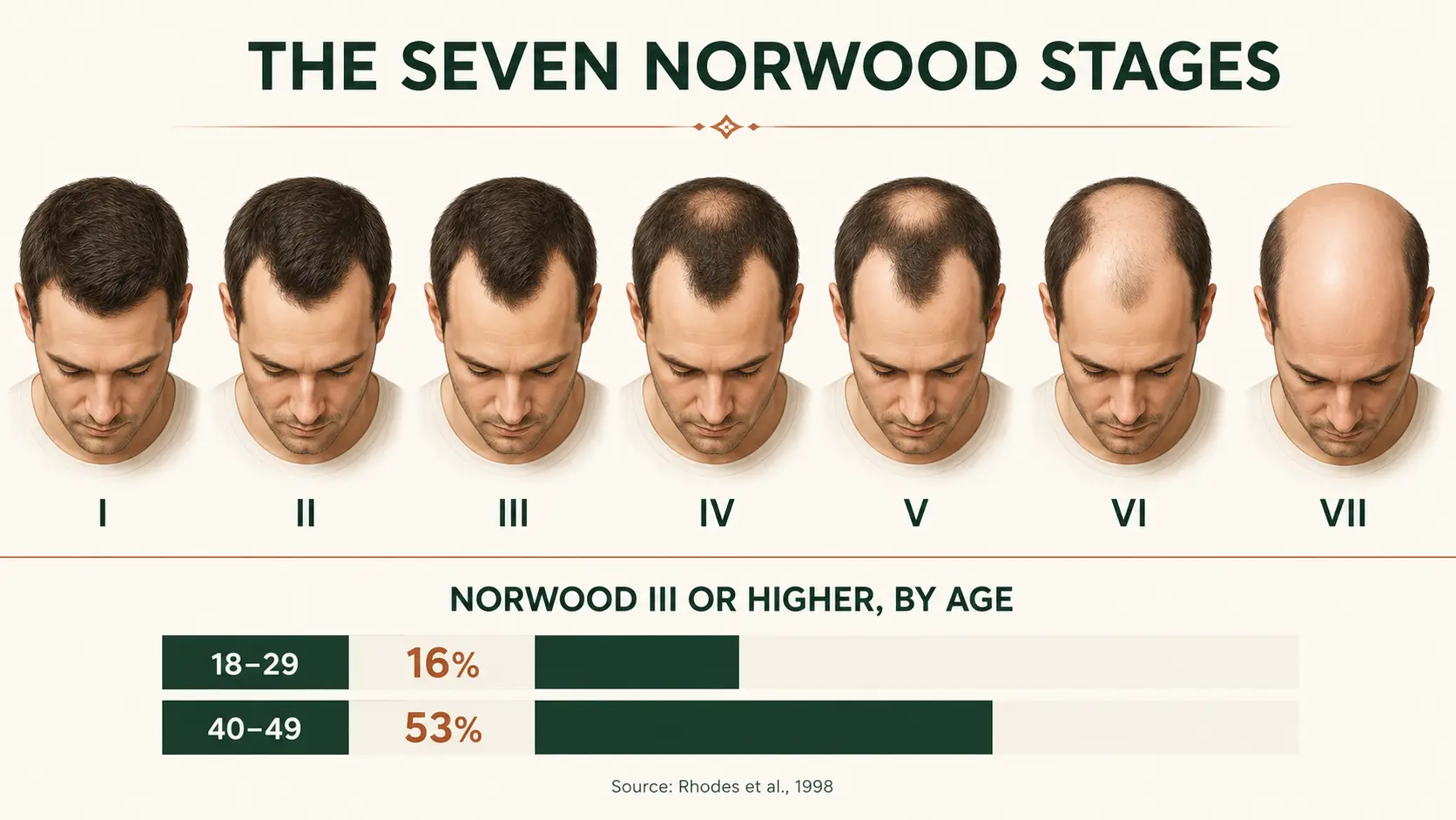
Norwood stages I to VII
Type I: no meaningful frontotemporal recession
The anterior hairline shows little or no recession. Hair transplantation isn't indicated for a normal Type I pattern. If there is shedding, diffuse thinning, scalp discomfort or a sudden change, I first look for a diagnosis that the Norwood diagram can't capture.
Type II: early recession or a mature hairline
The frontotemporal corners have moved back, but the recession remains limited. Some Type II hairlines are stable, age-appropriate mature hairlines. Others are an early frame in a moving sequence.
One photograph can't reliably separate the two. I compare older photographs, look for loss of density behind the line and examine whether the hairs at the edge are becoming finer. A low juvenile hairline shouldn't be recreated simply because the patient can still see where it used to sit.
Type IIA: the first anterior variant
In Type A patterns, the entire frontal edge moves backward more uniformly instead of leaving the familiar central forelock and deeper temporal recessions. Type IIA can therefore look less like an “M” and more like one broad, receded line.
The letter A doesn't automatically mean aggressive biology. Its surgical relevance comes from geometry: rebuilding a wide frontal band can consume far more grafts than filling two corners. In a young patient with active progression, that broad commitment deserves caution.
Type III: established frontal recession
Type III is generally the first Norwood category considered definite balding. The temporal recessions are deeper and the frontal outline becomes more clearly M-, U- or V-shaped.
This is also the stage at which many men start asking about surgery. The visible area may still be relatively limited, which can make a strong result possible, but the future pattern isn't always readable yet. Age, serial photographs and miniaturization matter more here than they do in a simple stage chart.
Type III Vertex: early crown involvement
Type III Vertex adds distinct loss at the crown while frontal recession remains no more advanced than Type III. This isn't a cosmetic footnote. The patient now has two recipient zones competing for the same finite donor supply.
In a younger patient, I become more conservative with the proposed hairline because crown loss may expand in surface area. Filling the front aggressively and treating the crown at the same time can use the reserve that a later mid-scalp transition will need. My separate guide to crown hair transplant planning explains why the whorl, surface area and future expansion change the calculation.
Type IIIA: frontal loss without a separate crown spot
Type IIIA continues the Type A path. The frontal hairline has receded farther as one broad front, while a separate vertex bald spot is absent.
The practical risk is easy to misunderstand. A visible crown spot may be absent, but a low new hairline across a wide forehead can still demand a large number of grafts. I need to know whether that frontal edge has been stable and whether the mid-scalp behind it is miniaturizing before deciding how much to rebuild.
Type IV: separate frontal and crown zones
Frontal recession is more advanced and vertex loss may be present, with a band of hair still separating the two areas. The pattern is often easier to recognize, but the bridge between the front and crown can be more important than either bald area.
If that bridge is miniaturizing, today's two separate zones may become one larger area. A plan that spends heavily on both ends can leave too little hair for the middle.
Type V: the bridge narrows
The frontal and vertex areas enlarge and the band between them becomes narrower or sparser. Donor demand now rises faster than the Norwood number suggests because the recipient surface is becoming continuous.
At this stage I usually prioritize facial framing and mid-scalp continuity. Crown density may need to remain lighter, be delayed or be omitted, depending on the donor assessment.
Type VI: the front and crown have joined
The bridge has largely disappeared, leaving one extensive area from the frontal scalp to the crown. Surgery can still be worthwhile in selected patients, but “full coverage” and “full density” become very different promises.
A retrospective series of 820 men with Norwood V–VII loss described the central problem as a donor–recipient mismatch. At 12 months, 94% of patients were satisfied with the result, and 62% still wanted another sitting for more coverage or density. [12] Both figures belong in the same sentence. Advanced cases can go well and still leave the patient wanting more, which is exactly why the cosmetic goal should be defined before harvesting begins.
Type VII: extensive loss with a narrow remaining rim
Only a relatively narrow horseshoe-shaped band remains at the sides and back. The donor may also show miniaturization, retrograde thinning or a boundary that is less stable than it first appears.
Some Type VII patients can achieve useful facial framing. Others don't have enough reliable donor hair to justify surgery. I would rather decline a case than create a thin transplanted island surrounded by future loss or leave the donor visibly depleted.
Norwood III vs III Vertex vs IIIA
These three labels can sit beside one another on a chart while describing very different surgical problems.
| Pattern | What is visibly different? | Main planning question | Common mistake |
|---|---|---|---|
| Norwood III | Deep temporal recession with an M-, U- or V-shaped frontal outline | Is the loss stable enough to rebuild the frame conservatively? | Treating a young patient's current corners as the final boundary of loss |
| Norwood III Vertex | Type III-or-less frontal recession plus a distinct crown area | How much donor reserve must be protected for two expanding zones? | Lowering the hairline and densely filling the crown in the same plan without budgeting for the mid-scalp |
| Norwood IIIA | The whole frontal line has moved back more uniformly; no separate vertex spot | How wide is the frontal recipient area, and is the hair behind it stable? | Assuming “no crown spot” means low donor demand |
The age-stage planning matrix
Age doesn't make somebody suitable or unsuitable by itself. It changes how much uncertainty remains and how long the donor plan must continue to make sense. The matrix below summarizes my usual planning bias before an individual examination.
| Age and stage | Progression uncertainty | Typical first response | Hairline and donor approach |
|---|---|---|---|
| 24, Norwood III | High | Confirm diagnosis, compare serial photographs, discuss evidence-based medical treatment and observe when the pattern is moving | Avoid an adolescent-height hairline; preserve reserve for loss behind it |
| 24, Norwood III Vertex | Very high | Assess both frontal and crown miniaturization; crown surgery often waits | Protect donor supply for a potentially expanding mid-scalp and crown |
| 28, Norwood V | Very high donor-demand risk | Stabilization and detailed donor mapping before any promise; some patients should be declined | Mature frontal design, staged priorities and explicit acceptance that the crown may remain light |
| 35–44, Norwood III–IV | Moderate and highly history-dependent | Compare at least two time points and examine miniaturization | Individualize the hairline; allocate density where it changes appearance most |
| 45, Norwood III | Often lower if photographs show years of stability | Surgery may be more predictable after diagnosis and donor assessment | A conservative but refined frontal restoration may be reasonable |
| 55, Norwood VI | Pattern may be readable, but recipient demand remains high | Measure the reliable donor and agree on the coverage priority | Mature framing and strategic coverage; original density isn't a realistic target |
This isn't a candidacy calculator. A stable 28-year-old with an excellent donor can be safer to plan than a 45-year-old with diffuse donor miniaturization. The useful comparison is always stage × age × rate of change × donor quality.
What the scale leaves out
Donor density and lifetime supply
The Norwood stage describes the recipient pattern. It says nothing about the amount of hair that can be moved safely.
In a 2019 cross-sectional study of 580 Indian men aged 24–45 with Norwood III or greater loss, mean scalp donor density was 78.2 follicular units/cm², mean hair density was 141.5 hairs/cm² and the average follicular unit contained 1.81 hairs. [8] Those are cohort averages, not a promise to an individual patient. The study is useful precisely because it shows what the Norwood number omits: even patients in the same grade can have materially different donor resources.
I assess density, hairs per follicular unit, caliber, miniaturization, the boundaries of the safe donor area, previous harvesting and signs of retrograde or diffuse thinning. An FUE plan should begin with the donor, not with a target graft quote. You can read more about that approach on the FUE hair transplant page.
Hair caliber, curl and scalp contrast
Caliber and contrast are often confused, so it is worth separating them:
- Thicker hair covers more surface area than fine hair.
- Wavy or curly hair usually creates more visual bulk than straight hair at the same numerical density.
- Low hair-to-scalp contrast makes gaps less visible. Light hair on light skin is a common example.
- High contrast exposes the scalp more clearly. Fine, dark hair on light skin is one of the most demanding combinations.
The most favorable combination is thick hair with low contrast and useful texture. One of the hardest is fine, straight, dark hair against a light scalp. Two Norwood IV patients can therefore receive the same number of grafts and look very different. [11,13]
Miniaturization and rate of change
The scale records an outline. It doesn't show how many apparently present hairs are already becoming thinner and shorter.
Trichoscopy can help assess hair-shaft diversity, miniaturization, donor density and follicular-unit characteristics before surgery. [10] Serial photographs add the time dimension. A standardized image from three years ago can sometimes tell me more about risk than a beautifully lit photograph taken yesterday.
Scalp health and the diagnosis itself
Redness, scale, pustules, loss of follicular openings, burning or an unusual distribution can change the diagnosis and the surgical timing. Active disease matters more than a convenient Norwood label.
Mild, controlled seborrheic dermatitis doesn't automatically exclude surgery. Scarring alopecias such as frontal fibrosing alopecia (FFA) or central centrifugal cicatricial alopecia (CCCA) require a completely different level of caution. My guide to scalp inflammation and hair transplantation explains those distinctions in more detail.
Why people often grade themselves incorrectly
Family photographs are useful, but they aren't destiny. A young patient may zoom into an uncle's wedding photograph and ask, “Will I become a Norwood VII?” The honest answer is that family history changes suspicion, not certainty. I make a safer plan by combining that history with the patient's own rate of change, examination and response to treatment.
Research supports the limits of self-grading. In one study, two trained observers achieved 85.4% exact agreement (κ = 0.828), while the men's exact agreement with the observers was only 48.5–55.8%. [5] Another study involving dermatologists and residents found unsatisfactory inter-rater agreement for the full Hamilton–Norwood scale (ICC 0.63–0.68) and poor repeatability. [4]
Those results aren't truly contradictory. Agreement changes with training, image quality, the number of categories and the way borderline patterns are handled.
I use the Norwood number as a shared description, not as a precise measurement.
When BASP is more useful
The BASP system was introduced in 2007 as a classification that could describe pattern hair loss in both men and women. It separates the anterior hairline shape into four basic types — L, M, C and U — and then records frontal and vertex density as the specific F and V types. [6]
That structure can describe combinations the Norwood scale handles poorly, including diffuse thinning and patterns that don't follow a simple front-and-crown sequence. In a comparison involving eight hair specialists, 17 dermatology residents and 15 general physicians grading 100 sets of photographs, BASP showed better reproducibility and repeatability than Norwood–Hamilton. [7]
For women, I don't force a male sequence onto a different pattern. Ludwig or BASP may be more informative, and the diagnostic workup is often different. The female hair transplant page explains why diffuse thinning, donor stability and the cause of loss need separate consideration. The same caution applies to transgender hairline planning, where the goal and the expected future pattern may both differ from the sequence the Norwood diagrams describe.
How I plan surgery beyond the number
I decide which area deserves the donor first
A graft creates different cosmetic value depending on where it is placed. The frontal frame usually changes the face more than the same number dispersed across a large crown. In advanced loss, spreading grafts evenly over every empty centimeter can create weak coverage everywhere.
The allocation needs to be agreed before surgery: frontal hairline, frontal third, mid-scalp, crown or a staged combination. The Norwood stage helps define the map; the priority defines the plan.
I design for the patient's future face
A hairline that looks impressive at 24 can look misplaced at 44 if the native hair behind it disappears. Height, temporal recession, shape, density gradient and the patient's facial proportions all need to age together.
This doesn't mean every young patient receives the same high line. It means I won't use a low hairline to solve today's anxiety while leaving tomorrow's donor problem unanswered.
I protect donor capacity for native hair that may be lost later
Hair transplantation redistributes follicles. It doesn't stop androgenetic alopecia in the surrounding native hair. International expert consensus emphasizes appropriate candidate selection and adequate medical treatment of alopecia before transplantation. [9]
Medication is an individual medical decision, not a condition I hide in the small print. Finasteride, dutasteride or minoxidil may help selected patients preserve miniaturizing hair, but risks, contraindications and long-term adherence need a real discussion. A patient who prefers to start with something “natural” can read my evidence review of rosemary oil for hair loss; it also explains why limited evidence shouldn't be confused with control of progressive androgenetic alopecia.
I keep the graft estimate separate from the sales number
Graft estimates depend on the area in square centimeters, planned density, existing native hair, caliber, hairs per graft and donor safety. A stage label can't calculate all of that.
This is why a larger quotation isn't automatically a better plan. If two clinics give different numbers, ask which zones each number covers, what density is planned, how the donor was measured and what reserve remains for a second procedure.
When another condition resembles a Norwood pattern
The Norwood Scale is intended for male pattern hair loss. A receded or thin area can have another cause, including traction alopecia, alopecia areata, frontal fibrosing alopecia, lichen planopilaris, central centrifugal cicatricial alopecia, telogen effluvium or a diffuse unpatterned process.
Warning signs include:
- sudden or patchy loss
- burning, pain or marked itching
- redness, pustules or heavy scale
- shiny skin or loss of visible follicular openings
- broken hairs or a distribution linked to tension
- diffuse thinning through the supposed donor area
If the diagnosis is wrong, the Norwood stage is irrelevant.
The bottom line
The Norwood Scale is useful because it gives patients and clinicians a common visual language. Its limits are just as important as its stages.
I don't plan a transplant from one diagram. I plan from the patient's age, serial photographs, miniaturization, donor measurements, hair characteristics, scalp health, medical history and tolerance for future uncertainty. The same Norwood number can justify surgery, observation, medical treatment or a decision not to operate.
Some of that medical history changes the operation itself rather than the design. A condition such as epilepsy, for example, affects the anesthetic plan and the timing far more than it affects the hairline.
The goal is a result that fits the patient today, still makes sense a decade later and never advertises the fact that surgery happened.
Frequently asked questions
What is the Norwood Scale?
How can I find my own Norwood stage?
How many grafts does Norwood III need?
Is Norwood II hair loss or a mature hairline?
What is the difference between Norwood III and III Vertex?
What do Norwood IIA and IIIA mean?
Can two people at the same Norwood stage need different surgery?
Can a Norwood stage go backward?
How quickly does hair loss move through Norwood stages?
When is the right time to have a hair transplant?
Does the Norwood Scale apply to women?
Which Norwood stage is best for a hair transplant?
Can a Norwood VI or VII patient still have surgery?
Does a higher Norwood stage always mean more grafts?
— Dr. Mesut Demir
Want an assessment that goes beyond a stage number?
Send clear photographs of the front, both temples, top, crown and donor area. Include your age, when you first noticed change, current treatment and one or two older photographs if available. I can then explain the likely pattern, what still needs to be confirmed, whether the timing looks sensible and which areas deserve priority. The purpose is a realistic long-term plan, not the largest graft quote.
Request a case review- Hamilton JB. Patterned loss of hair in man: types and incidence. Annals of the New York Academy of Sciences. 1951;53(3):708–728. doi:10.1111/j.1749-6632.1951.tb31971.x
- Norwood OT. Male pattern baldness: classification and incidence. Southern Medical Journal. 1975;68(11):1359–1365. doi:10.1097/00007611-197511000-00009
- Rhodes T, Girman CJ, Savin RC, et al. Prevalence of male pattern hair loss in 18–49 year old men. Dermatologic Surgery. 1998;24(12):1330–1332. doi:10.1111/j.1524-4725.1998.tb00009.x
- Guarrera M, Cardo P, Arrigo P, Rebora A. Reliability of Hamilton–Norwood classification. International Journal of Trichology. 2009;1(2):120–122. doi:10.4103/0974-7753.58554
- Taylor R, Matassa J, Leavy JE, Fritschi L. Validity of self-reported male balding patterns in epidemiological studies. BMC Public Health. 2004;4:60. doi:10.1186/1471-2458-4-60
- Lee WS, Ro BI, Hong SP, et al. A new classification of pattern hair loss that is universal for men and women: basic and specific (BASP) classification. Journal of the American Academy of Dermatology. 2007;57(1):37–46. doi:10.1016/j.jaad.2006.12.029
- Hong H, Ji JH, Lee Y, Kang H, Choi GS, Lee WS. Reliability of the pattern hair loss classifications: a comparison of the BASP and Norwood–Hamilton classifications. Journal of Dermatology. 2013;40(2):102–106. doi:10.1111/1346-8138.12024
- Chouhan K, Kota RS, Kumar A, Gupta J. Assessment of safe donor zone of scalp and beard for follicular unit extraction in Indian men: a study of 580 cases. Journal of Cutaneous and Aesthetic Surgery. 2019;12(1):31–35. doi:10.4103/JCAS.JCAS_142_18
- Vañó-Galván S, Bisanga CN, Bouhanna P, et al. An international expert consensus statement focusing on pre- and post-hair transplantation care. Journal of Dermatological Treatment. 2023;34(1):2232065. doi:10.1080/09546634.2023.2232065
- Issa NT, Tosti A. Trichoscopy for the hair transplant surgeon — assessing for mimickers of androgenetic alopecia and preoperative evaluation of donor site area. Indian Journal of Plastic Surgery. 2021;54(4):393–398. doi:10.1055/s-0041-1739245
- Goldin J, Zito PM, Raggio BS. Hair transplantation. StatPearls, NCBI Bookshelf. Updated 2026. NBK547740
- Chouhan K, Roga G, Kumar A, Gupta J. Approach to hair transplantation in advanced grade baldness by follicular unit extraction: a retrospective analysis of 820 cases. Journal of Cutaneous and Aesthetic Surgery. 2019;12(4):215–222. doi:10.4103/JCAS.JCAS_173_18
- Khanna M. Hair transplantation surgery. Indian Journal of Plastic Surgery. 2008;41(Suppl):S56–S63. PMID 20174544
- British Association of Dermatologists. Male pattern hair loss (androgenetic alopecia) — patient information leaflet.